May Thurner Syndrome Radiology
May thurner syndrome radiology. May and Thurner in 1957 described left common iliac vein intraluminal bands resulting from mechanical obstruction by the right common iliac artery. This single-center retrospective review identified 6 patients n 6 100 female treated by endovascular therapy for May-Thurner syndrome from June 2013 to September 2015. To evaluate the feasibility of endovascular techniques in treating venous outflow obstruction resulting from compression of the iliac vein by the iliac artery of.
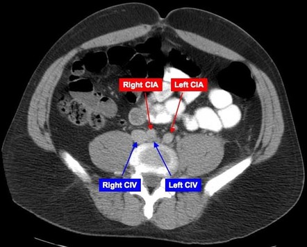
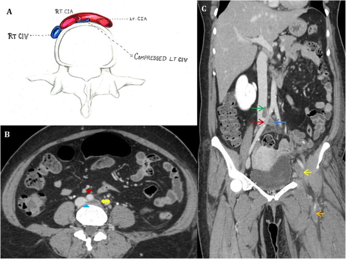
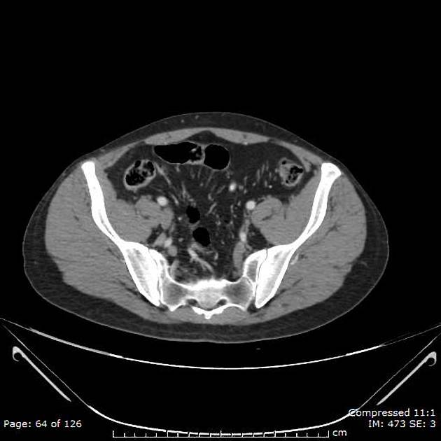
May-Thurner syndrome refers to a chronic compression of the left common iliac vein CIV against the lumbar vertebrae by the overlying right common iliac artery CIA with or without deep venous thrombosis 1 article features images from this case. The most common symptoms are leg swelling and discomfort. May-Thurner syndrome is always the working diagnosis when a patient presents with unilateral left lower limb swelling without signs of infection.
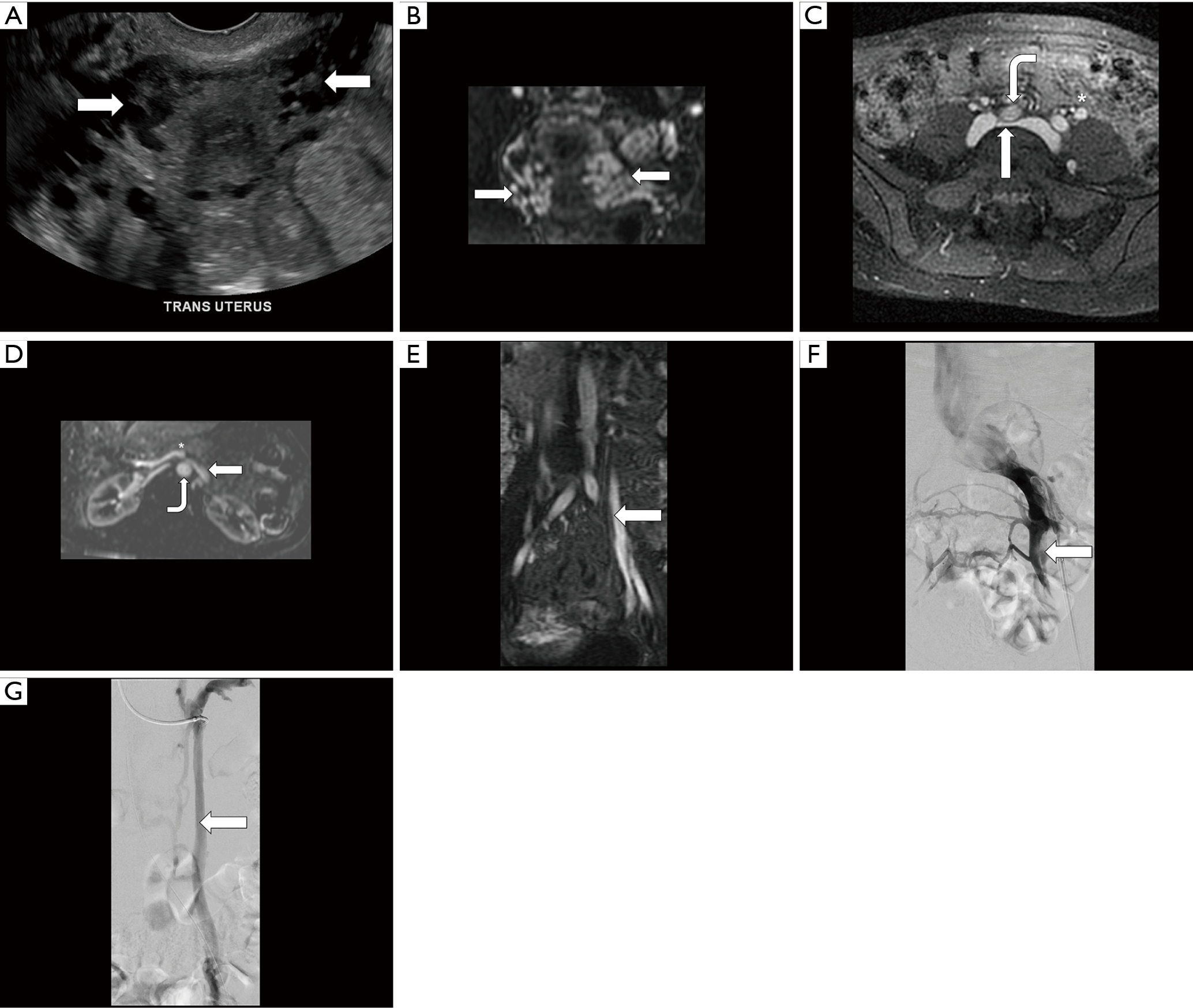
The typical patient with May-Thurner syndrome has left lower extremity pain and swelling resulting in varicosities phlebitis deep venous thrombosis venous stasis dermatitis and pulmonary embolism. When symptomatic such compressions can result in a variety of uncommon syndromes in the abdomen and pelvis including median arcuate ligament syndrome MALS May-Thurner syndrome nutcracker syndrome superior mesenteric artery SMA syndrome UPJ obstruction ovarian vein syndrome OVS and other forms of ureteral compression Table. 5 Patel NH Stookey KR Ketcham DB Cragg AH.
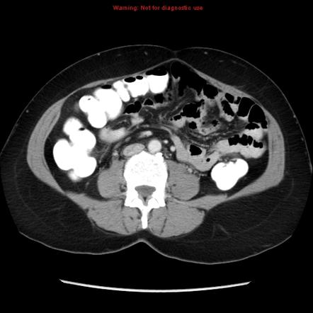
1 article features images from this case. The right iliac vein is patent. All three patients had a spur May-Thurner syndrome of the left common iliac vein at the level of the crossover position of the right common iliac artery.
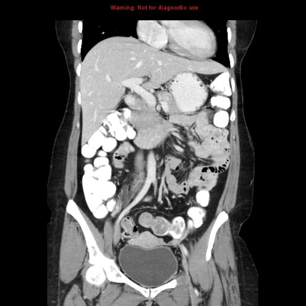
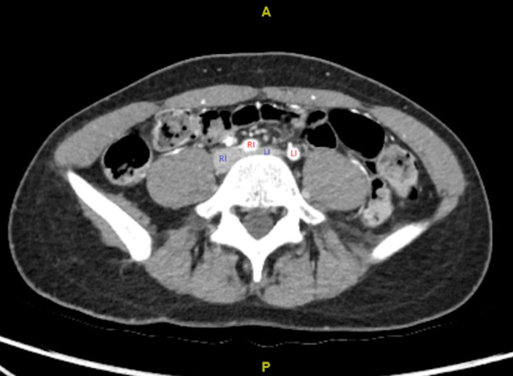
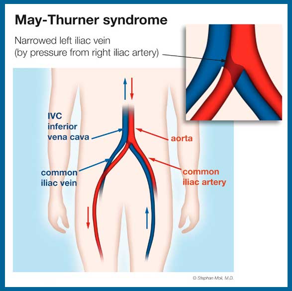
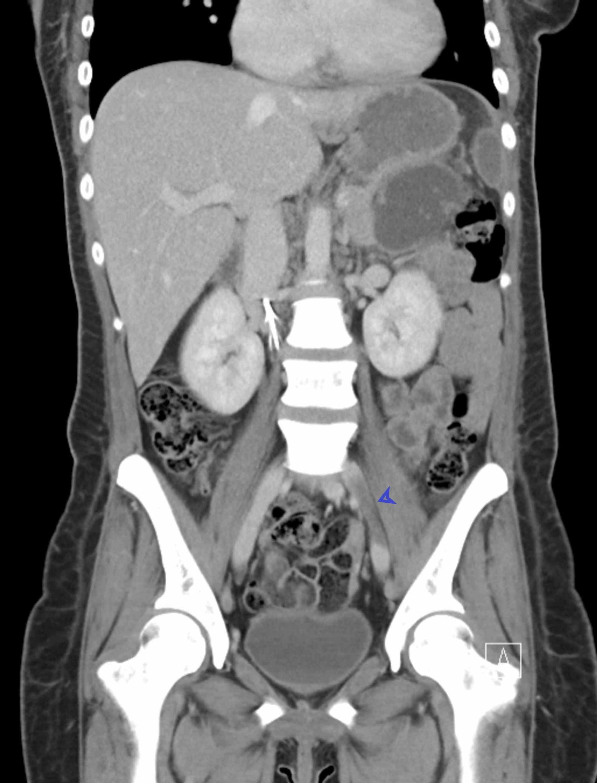
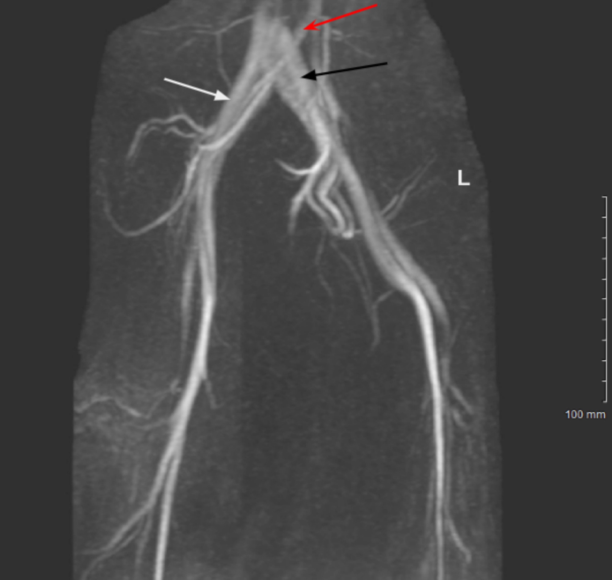
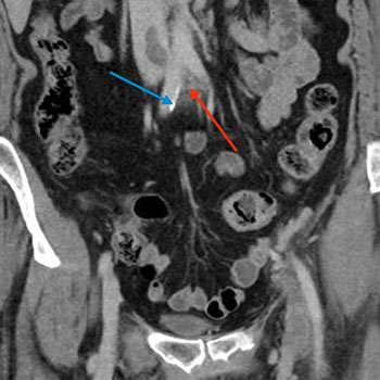
The left iliac vein is partially thrombosed. The vertical linear defect of the left common iliac vein is consistent with May-Thurner syndrome. About May-Thurner Syndrome May-Thurner syndrome is a pelvic condition in which the right common iliac artery compresses the left iliac vein pressing it against the spine.
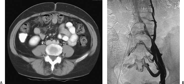
May Thurner Syndrome MTS is a condition in which the left iliac vein is compressed by the right iliac artery. May-Thurner syndrome can also cause development of extensive retroperitoneal and pudendal venous collaterals which shunt the distal iliofemoral venous system to the contralateral deep venous vessels Figs. Patients consisted of 3 African American 2 Caucasian and 1 Asian.
May-Thurner syndrome is deep venous thrombosis resulting from chronic compression of the left common iliac vein CIV against the lumbar vertebrae by the overlying right common iliac artery CIA. Three patients with long standing edema of the left leg were examined and venography and hemodynamic studies were performed.
The most common symptoms are leg swelling and discomfort.
This vein compression can lead to blood clots such as deep vein thrombosis. Both of these conditions create alternative venous drainage to counteract the venous stasis preventing. All three patients had a spur May-Thurner syndrome of the left common iliac vein at the level of the crossover position of the right common iliac artery. May-Thurner syndrome is always the working diagnosis when a patient presents with unilateral left lower limb swelling without signs of infection. Mean age was 5350 831 years range. May Thurner Syndrome MTS is a condition in which the left iliac vein is compressed by the right iliac artery. May-Thurner syndrome is deep venous thrombosis resulting from chronic compression of the left common iliac vein CIV against the lumbar vertebrae by the overlying right common iliac artery CIA. D After placement of a 10- 40-mm Wallstent across the left common iliac vein there is free flow across the common iliac vein with resolution of collateral flow. MDCT is useful for fast comprehensive evaluation of the vascular system to determine whether May-Thurner syndrome or an alternative condition is present.
To evaluate the feasibility of endovascular techniques in treating venous outflow obstruction resulting from compression of the iliac vein by the iliac artery of. The typical patient with May-Thurner syndrome has left lower extremity pain and swelling resulting in varicosities phlebitis deep venous thrombosis venous stasis dermatitis and pulmonary embolism. The left iliac vein is partially thrombosed. This vein compression can lead to blood clots such as deep vein thrombosis. All three patients had a spur May-Thurner syndrome of the left common iliac vein at the level of the crossover position of the right common iliac artery. The vertical linear defect of the left common iliac vein is consistent with May-Thurner syndrome. 1 article features images from this case.













Posting Komentar untuk "May Thurner Syndrome Radiology"